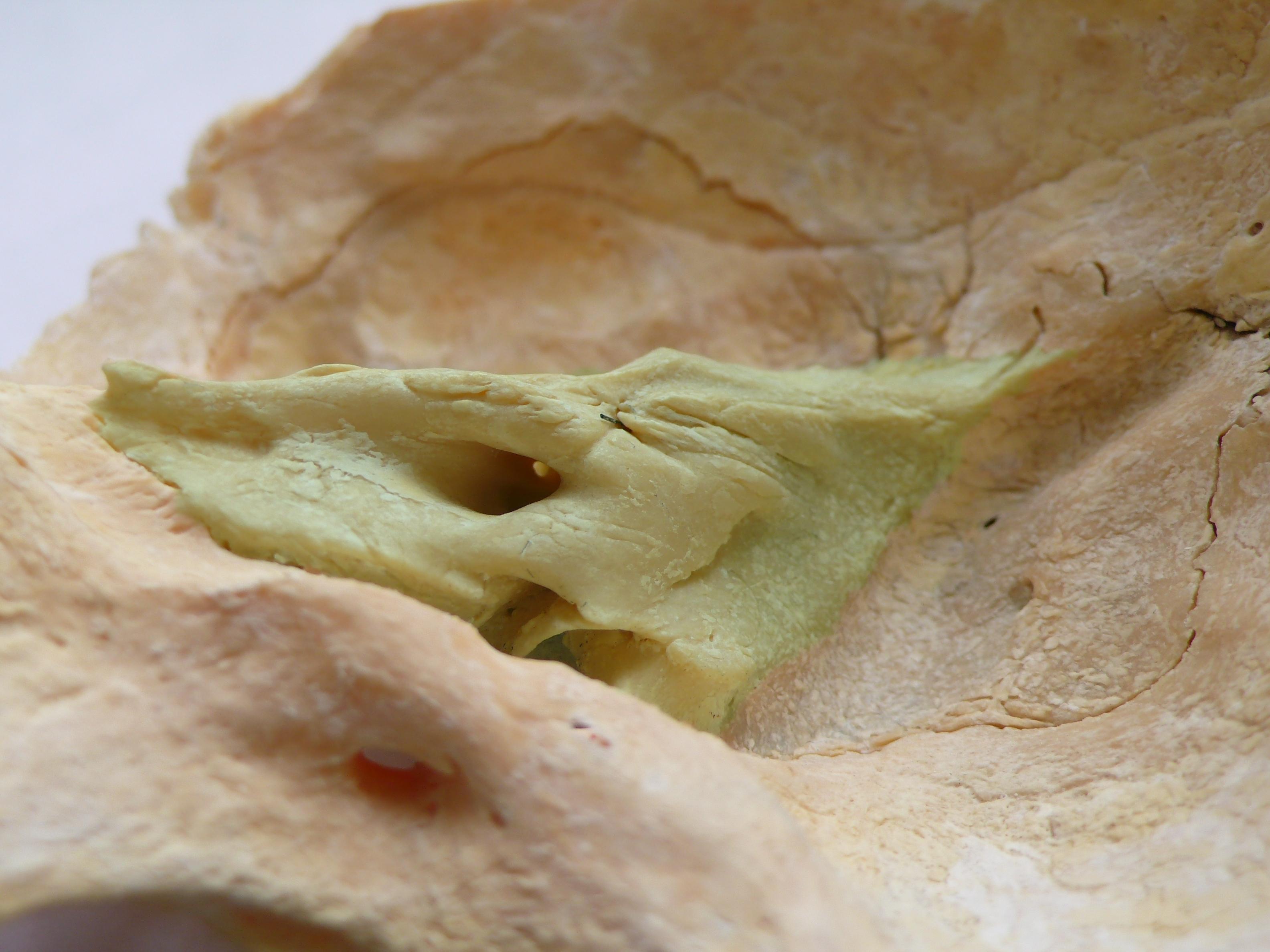
µCT scanning has become a valuable tool for documenting archaeological human remains before destructive sampling, especially when researchers work with the petrous portion of the temporal bone, one of the richest sources of ancient DNA.
This study tests whether routine µCT imaging actually damages ancient DNA preservation in archaeological petrous samples. Across 93 specimens, the authors do not find strong evidence that scanning systematically worsens key aDNA quality measures.
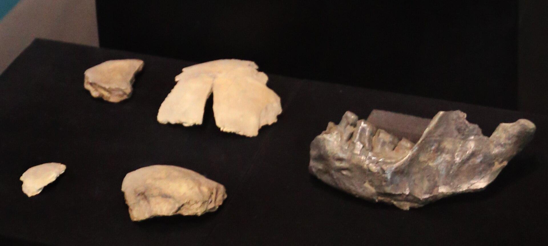
Why the petrous bone matters
The petrous bone is prized in archaeogenetics because its dense structure often preserves DNA better than most other skeletal elements. But that same importance creates a tension: imaging, osteological study, and molecular sampling all compete for access to a limited and valuable piece of anatomy.
What the study found
By comparing scanned and unscanned material, the researchers show that standard µCT workflows do not appear to cause major losses in endogenous DNA, coverage, or other core sequencing outcomes. That is encouraging for archaeologists who want better anatomical documentation without automatically compromising future genetic work.
A more sustainable workflow
Beyond the preservation question, the paper proposes a practical workflow that better balances imaging, osteobiography, and destructive sampling. The broader message is that documentation and biomolecular analysis do not have to be treated as enemies if collections are handled carefully and in the right order.
Source article: https://pmc.ncbi.nlm.nih.gov/articles/PMC13075688/


Comments